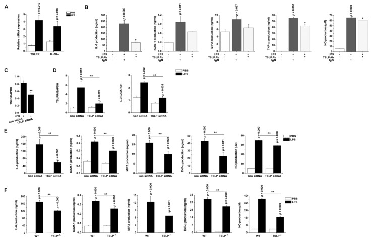
Figure 5.
TSLP-dependent proinflammatory environment predisposes to the development of sepsis. (A) The mRNA expression of TSLPR and IL-7Rα were analyzed in RAW 264.7 cells by real-time PCR. (B) RAW 264.7 cells were stimulated with LPS (0.1 μg/mL) in the absence or presence of neutralizing anti-TSLP antibody (5 µg/mL) or nonimmune IgG (5 µg/mL) for 24 h. # p < 0.05 vs. LPS stimulation. RAW 264.7 cells were transfected with scramble control siRNA or TSLP-specific siRNA. After LPS stimulation, the mRNA expression levels of (C) TSLP, (D) TSLPR, and IL-7Rα were analyzed in the transfected cells by real-time PCR. (E) The transfected RAW 264.7 cells were stimulated with LPS for 24 h. ** p < 0.05 vs. Con siRNA transfection and LPS stimulation. (F) The peritoneal macrophages isolated from TSLP−/− mice were stimulated ex vivo with LPS for 24 h. Each production was analyzed by ELISA. Nitric oxide concentration was measured by the Griess method. Data are representative of three independent experiments (n = 5/group). A p value indicates the significant difference between PBS and LPS. ** p < 0.05 vs. LPS-stimulated WT macrophages. PBS, phosphate-buffered saline; LPS, lipopolysaccharide; TSLP, thymic stromal lymphopoietin; Ab, antibody; Con, control; siRNA, small interfering RNA; ICAM-1, intercellular adhesion molecule-1; MIP2, macrophage inflammatory protein 2; NO, nitric oxide; WT, wild-type.

