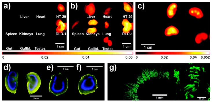
Figure 5.
Ex vivo fluorescence images of organs and tumors: 2D-fluorescence reflectance imaging images of the model drug DY-676 (a) and HPMA copolymer (b) of mouse that was treated with star-like HPMA copolymer (polymer B); distribution of the model drug in kidneys 24 h after intravenous administration; left: placebo, middle: star-like HPMA, right: linear HPMA (c); pseudo-colored fluorescence images of kidney slices 24 h after i.v. injection—model drug: blue, HPMA polymer: yellow (d–f) (linear HPMA: d and e, star-like HPMA: f); Confocal microscopic images of the model drug distribution in the kidney 24 h after i.v. injection of 1.5 mg linear HPMA (polymer A) (g). Reprinted with permission from [31], Copyright [2012], American Chemical Society.

