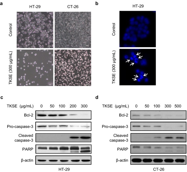
Fig. 3.
Effect of TKSE on colorectal cancer cell apoptosis. a HT-29 and CT-26 cells were treated with 300 µg/mL TKSE for 24 h. Representative photographic images are shown (Original magnification ×100). Scale bar = 100 µm. b The induction of apoptosis by TKSE (300 µg/mL) was assessed by DAPI staining (×100). The expressions of apoptosis-related proteins were assayed by western blotting HT-29 (c) or CT-26 (d) cells treated with TKSE at the indicated doses for 24 h

