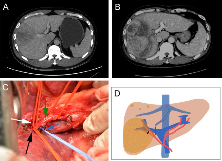
Fig. 2.
Preoperative findings and operative schema of patient 3 during the first stage. a Preoperative contrast CT revealed a huge hepatocellular carcinoma in the right lobe of liver. b The contrast CT showed an obvious volume increase of the future liver remnant on the postoperative day 13. c After the extraparenchymal separation, the main branch of right PV (blue arrow), the main branch of right HA (green arrow), the secondary arterial branches of the right anterior lobe (white arrow), and the right posterior lobe (black arrow) were exposed. d Operative schema of the first stage. The main branch of right PV was ligated (green line); the arterial branch of the right posterior lobe was ligated (black line)

