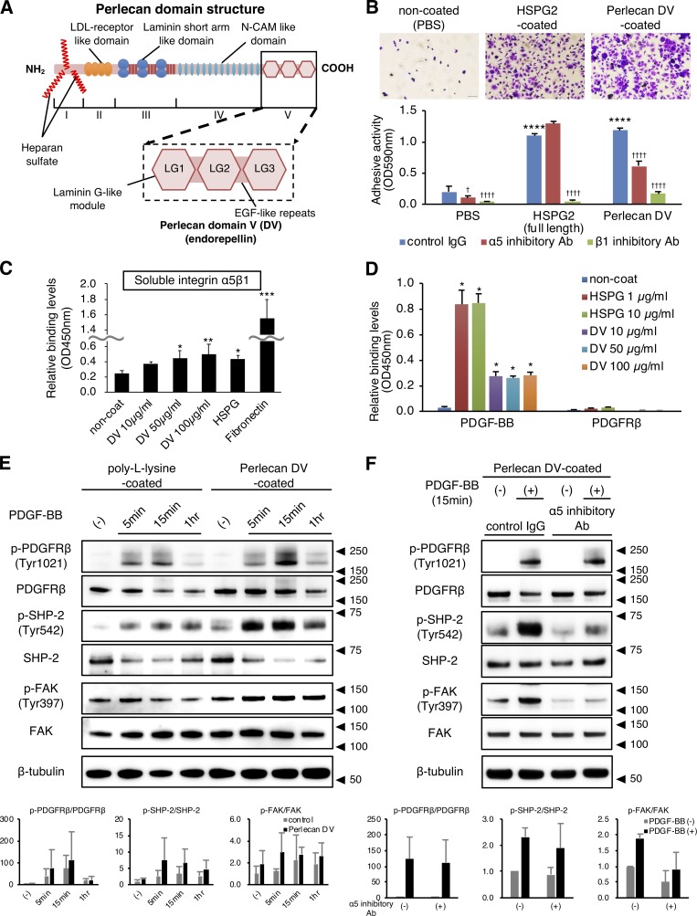
Figure 4.
Perlecan DV promotes PDGFRβ signaling through integrin α5β1. (A) Structure of perlecan. The five domains are numbered from the N terminus to the C terminus. Domain I contains the binding sites for heparan sulfate side chains. DV/endorepellin contains three globular domains that have homology to the laminin G module (LG) and that are each separated by epidermal growth factor (EGF)-like repeats (Noonan et al., 1991; Iozzo, 2005). (B) Representative images of brain pericytes attached to immobilized full-length perlecan (HSPG2, 10 µg/ml) or perlecan DV (500 nmol/liter; upper panels). Scale bar = 100 µm. The adhesion of pericytes to perlecan DV was inhibited by integrin function-blocking antibodies against integrins α5 (mAb16, 50 mg/ml) and β1 (mAb13, 50 mg/ml). Values are mean ± SD; n = 5; ****, P < 0.0001 versus PBS; †, P < 0.05; ††††, P < 0.0001 versus control IgG, one-way ANOVA followed by Tukey–Kramer’s HSD test. (C) Solid-phase binding assays of soluble integrin α5β1 (4 µg/ml) to the immobilized perlecan DV (10–100 µg/ml), full-length perlecan (HSPG2, 10 µg/ml), or fibronectin (10 µg/ml). Values are mean ± SD; n = 6; *, P < 0.05; **, P < 0.01; ***, P < 0.0001 versus noncoated, one-way ANOVA followed by Dunnett’s test. (D) Solid-phase binding assays of soluble PDGF-BB (0.5 µg/ml) or PDGFRβ (2 µg/ml) to the immobilized full-length perlecan (HSPG2) or perlecan DV. Values are mean ± SD; n = 3; *, P < 0.0001 versus noncoated, one-way ANOVA followed by Dunnett’s test. (E) The immunoblotting for the temporal profiles of p-PDGFRβ (Y1021)/PDGFRβ, p-SHP-2 (Y542)/SHP-2, and p-FAK (Y397)/FAK in pericytes. Cells were cultured on either poly-l-lysine (control, 15 µg/ml) or perlecan DV (500 nmol/liter). After serum starvation for 16 h, PDGF-BB (50 ng/ml) was added at the indicated time. A representative example of three independent experiments is shown (upper panels). The phosphorylated protein was quantitatively evaluated by densitometry, normalized with the total protein, and represented as the fold increase above the level obtained with control substrate without PDGF-BB (lower panels). Values are mean ± SD; n = 3. (F) The immunoblotting for p-PDGFRβ (Y1021)/PDGFRβ, p-SHP-2 (Y542)/SHP-2, and p-FAK (Y397)/FAK in pericytes under an inhibitory antibody for integrin α5. The cells were cultured on perlecan DV, serum-starved, pretreated with either control IgG or mAb16 (50 µg/ml) for 1 h, and incubated with PDGF-BB (20 ng/ml) for 15 min. A representative example of three independent experiments is shown (upper panels). The data represent the fold increase above the level obtained with control IgG treatment without PDGF-BB (lower panels). Values are mean ± SD; n = 3.

