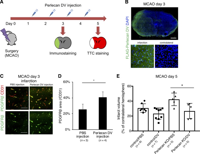
Figure 7.
Perlecan DV may promote the repair process of the BBB in ischemic stroke. (A) Perlecan DV was intraperitoneally injected daily for two or four consecutive days starting 24 h after MCAO. The mice were sacrificed at PSD 3 after MCAO for immunostaining or at PSD 5 for TTC staining. (B) Representative images of the immunostaining for FLAG. The administered recombinant perlecan DV with 3×FLAG tag was in infarct lesion at PSD 3 after MCAO. Scale bar = 1 mm or 50 µm. (C) Representative images of the immunostaining for PDGFRβ (green) and CD31 (red) at PSD 3 after MCAO, followed by perlecan DV injection in control mice. Perlecan DV–injected mice showed increased numbers of PDGFRβ-positive pericytes in the ischemic lesion at PSD 3. Scale bar = 100 µm. (D) PDGFRβ-positive areas were quantified and standardized by CD31-positive areas in the brain cortex. Values are mean ± SD; n = 3–4 per mice group; *, P < 0.05, unpaired t test. (E) The infarction volume in Perlecan KO mice at PSD 5, evaluated by TTC staining, was significantly smaller in the mice administered perlecan DV than in mice administered PBS-vehicle. Values are mean ± SD; n = 5–9 per mice group; *, P < 0.05, one-way ANOVA followed by Tukey–Kramer’s HSD test.

