Figure 3.
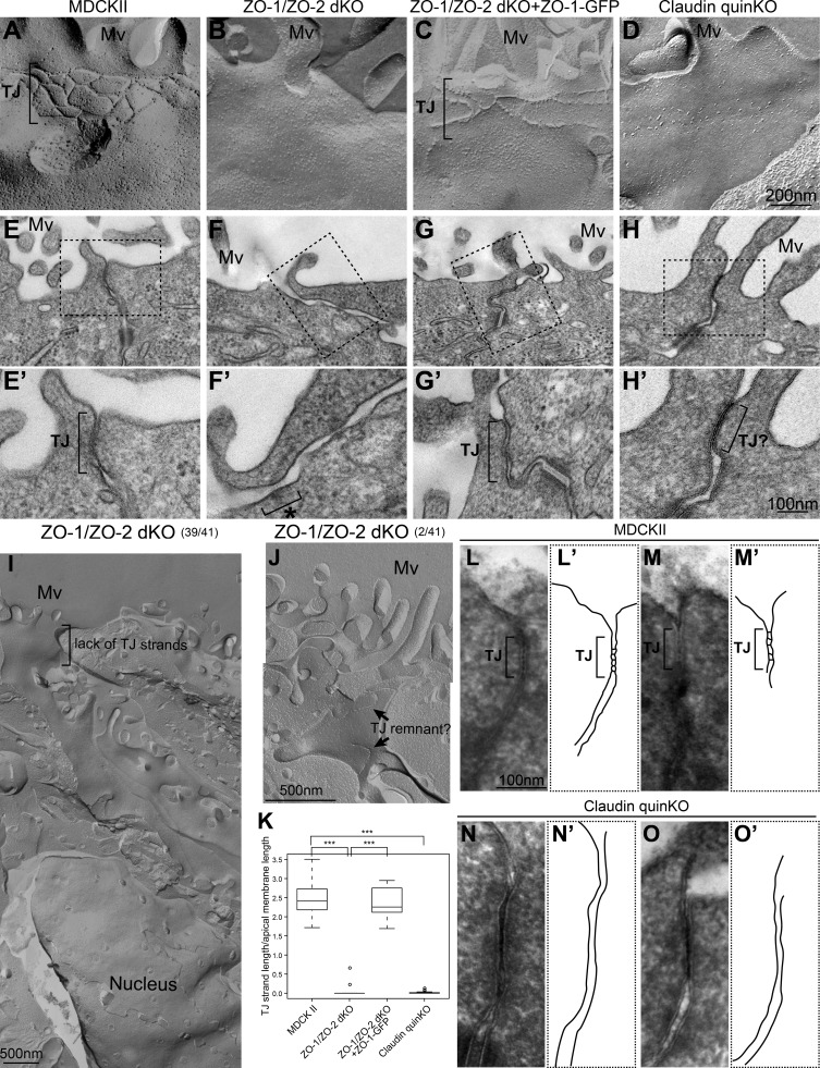
Claudins are required for TJ strand formation but not membrane appositions. (A–D) Freeze-fracture replica EM analyses. (A) TJ strands were observed beneath the apical microvilli in parental MDCK II cells. (B) TJ strands were not found in ZO-1/ZO-2 dKO cells. (C) Expression of ZO-1–GFP restored the TJ strands in ZO-1/ZO-2 dKO cells. (D) Claudin quinKO cells lacked TJ strands, but intramembrane particles were occasionally accumulated beneath the apical microvilli. (E–H) Transmission EM analyses of ultrathin sections. Black squares indicate the regions shown in high-magnification images. (E) TJs with membrane appositions were observed at the most apical cell junctions in MDCK II cells. (F) TJs were absent, and the intercellular space was widened in ZO-1/ZO-2 dKO cells. AJ-like structures associated with actin bundles were observed (asterisk). (G) Expression of ZO-1–GFP restored the formation of TJs in ZO-1/ZO-2 dKO cells. (H) TJ-like structures with membrane appositions were found in claudin quinKO cells. (I) Low-magnification view of a freeze-fracture replica from ZO-1/ZO-2 dKO cells. No TJ strands were found throughout the lateral plasma membrane. (J) An example of fragmented TJ strand-like structures in ZO-1/ZO-2 dKO cells. (K) Quantitation of TJ strand length normalized to the apical surface length of the corresponding fractured region. Graphs represent mean ± SD (n = 15 for MDCK II, n = 24 for ZO-1/ZO-2 dKO, n = 13 for ZO-1/ZO-2 dKO + ZO-1–GFP, n = 22 for claudin quinKO). ***, P < 0.0005, compared by t test. (L–O) TJ membrane kissing points were observed after ferrocyanide-reduced osmium/tannic acid/osmium postfixation. (L and M) TJ kissing points were observed in MDCK cells. (L′ and M′) Tracing of TJs. (N and O) Membranes were closely apposed to one another, but membrane kissing points were not observed in claudin quinKO cells. The most apical cell junctions with membrane appositions were observed. (N′ and O′) Tracing of TJ-like structures. Mv, microvilli. Scale bars: 200 nm (A–D); 100 nm (E–H); 500 nm (I and J); 100 nm (L–O).

