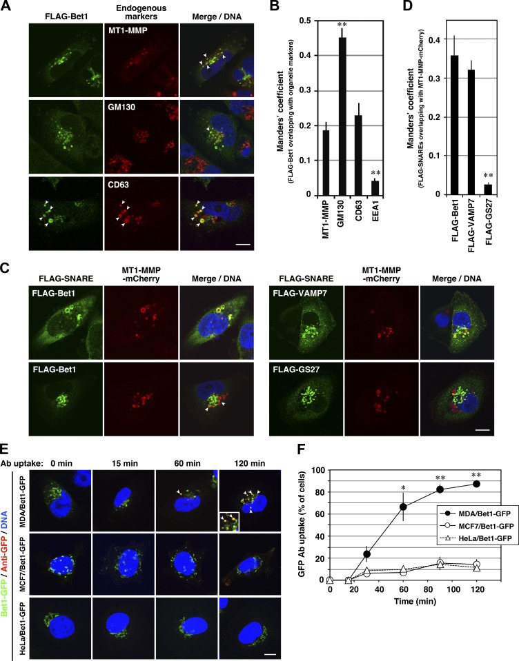
Figure 2.
Bet1 is localized in MT1-MMP–positive endosomes and reaches and is endocytosed from the plasma membrane. (A–D) Bet1 is colocalized with MT1-MMP in endosomal structures. MDA-MB-231 (A) and MDA-MT1-mCh (C) cells transiently expressing FLAG-Bet1 were fixed and then visualized with endogenous MT1-MMP, GM130, or CD63 (A) or MT1-MMP-mCherry (C) by confocal microscopy. In C, FLAG-VAMP7 and FLAG-GS27 were analyzed as FLAG-Bet1. Their colocalization was evaluated with the Manders’ overlap coefficient (B and D). (E) Bet1 reaches the plasma membrane and is endocytosed in MDA-MB-231 cells. MDA-MB-231, MCF7, and HeLa cells stably expressing Bet1-GFP were cultured in the complete medium containing anti-GFP antibodies for the indicated times, fixed, permeabilized, stained with secondary antibodies for anti-GFP, and then visualized by confocal microscopy. (F) Quantitation of the ratio of the cells that take up anti-GFP. Scale bar: 10 μm. *, P < 0.05; **, P < 0.01; vs. MT1-MMP in B; vs. FLAG-Bet1 in D; vs. MCF7/Bet1-GFP and HeLa/Bet1-GFP in F.

