Figure 7.
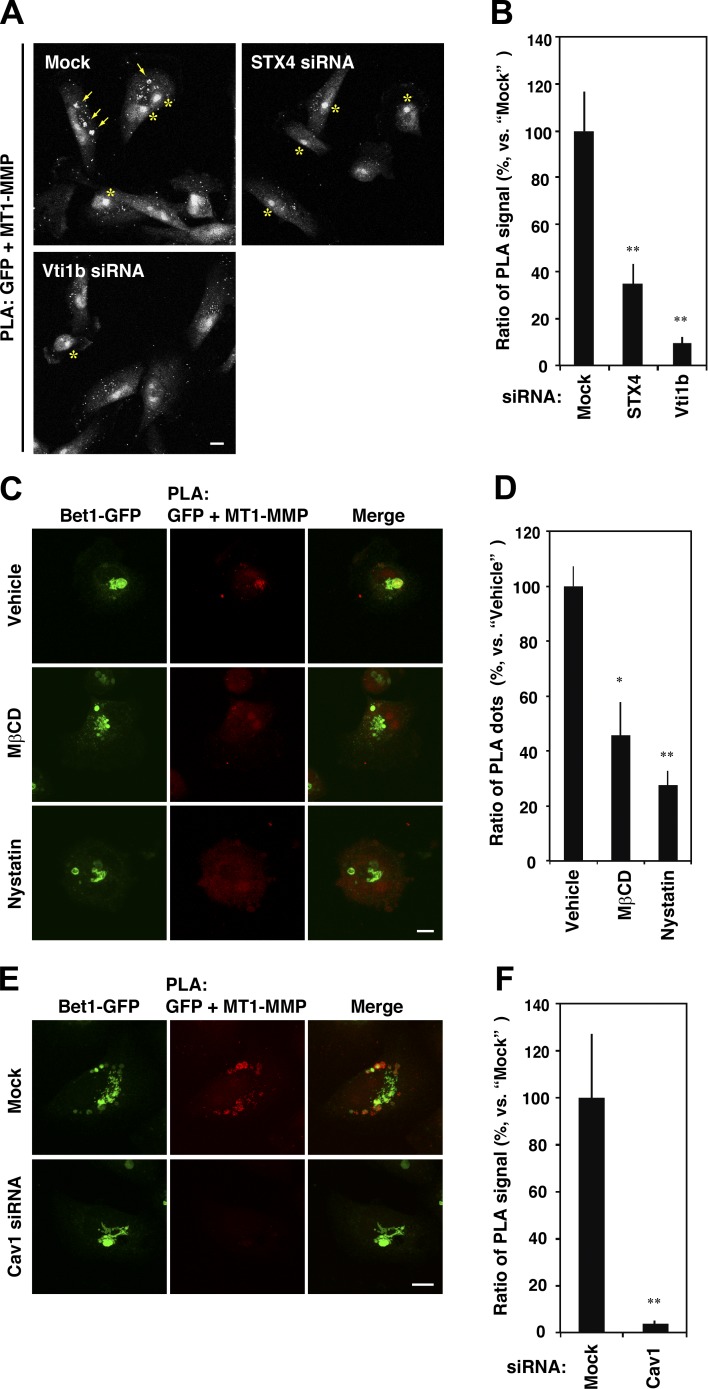
MT1-MMP is located in proximity to Bet1 in a raft-like, cholesterol-rich membrane domain. (A) Depletion of STX4 and Vti1b reduces the PLA signals between MT1-MMP and Bet1-GFP. MDA-MB-231 cells stably expressing Bet1-GFP were transfected with STX4 and Vti1b siRNAs, and the PLA was performed as in Fig. 6 E. Arrows indicate accumulated PLA signals in endosomes, whereas asterisks indicate nonspecific staining in nuclei. (B) Quantitation of PLA signals between MT1-MMP and Bet1-GFP as in A. (C) Cholesterol depletion weakens the interplay between MT1-MMP and Bet1. MDA-MB-231 cells stably expressing Bet1-GFP were spread on a fibronectin-coated coverslip for 7 h and then treated with 0.1% DMSO (vehicle), 5 mM methyl-β-cyclodextrin (MβCD) or 50 µg/ml nystatin for 30 min. After that, the cells were fixed and then subjected to PLA for MT1-MMP and Bet1-GFP. (D) Quantitation of PLA signals between MT1-MMP and Bet1-GFP as in C. (E) Depletion of Cav1 abolishes the proximity between MT1-MMP and Bet1-GFP. The experiments were performed as in A except for the use of Cav1 siRNA. (F) Quantitation of PLA signals between MT1-MMP and Bet1-GFP as in E. Scale bar: 10 μm. *, P < 0.05; **, P < 0.01; vs. mock in B and F; vs. vehicle in D.

