Figure 6.
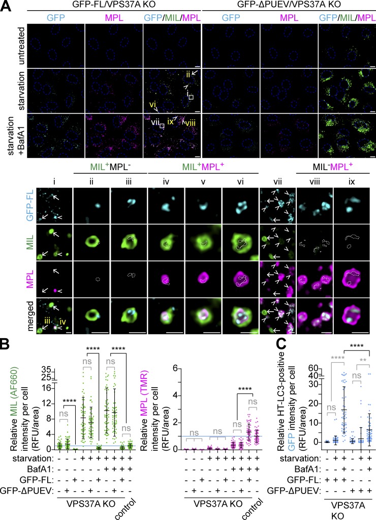
The PUEV domain of VPS37A is required for autophagosome completion. (A) HT-LC3–expressing U-2 OS cells with the indicated genotypes and transgenes were starved in the presence or absence of 100 nM BafA1 for 3 h and subjected to the HT-LC3 assay using AF660-conjugated MIL- and TMR-conjugated MPL. Representative confocal microscopy images from three independent experiments are shown. Magnified images in the boxed and arrow- or arrowhead-indicated areas are shown in the lower panels. Arrows and arrowheads indicate GFP-VPS37A–positive phagophores (MIL+MPL−) and autophagic vacuoles (MIL+MPL+ or MIL−MPL+), respectively. In the magnified images, dashed-line areas indicate the location of GFP-FL signals. The scale bars represent 10 µm in the main panels and 1 µm in the magnified images. (B and C) The cytoplasmic fluorescence intensities of MIL and MPL (B), and MIL− and/or MPL+ GFP-VPS37A (C) in each cell in A were quantified and normalized to the respective mean fluorescence intensities of control Cas9-expressing U-2 OS cells starved in the presence of BafA1 (n = 55). Statistical significance was determined by Kruskal–Wallis one-way ANOVA on ranks followed by Dunn’s multiple comparison test. All values are mean ± SD. **, P ≤ 0.01; ****, P ≤ 0.0001; ns, not significant.

