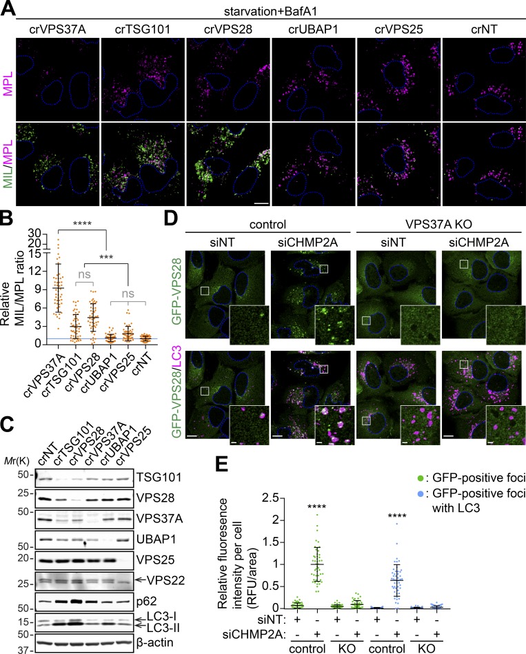
Figure 8.
VPS37A is responsible for ESCRT-I localization to the phagophore. (A–C) Three lentiviruses encoding different gRNAs against a gene were pooled and transduced into the HT-LC3–expressing U-2 OS cell for 2 d. Cells were then selected with 1 µg/ml puromycin for 3 d, cultured another 2 d in complete medium, and subjected to IB using the indicated antibodies (C) or starved in the presence of 100 nM BafA1 for 3 h followed by the HT-LC3 assay (A). In A, representative confocal images from three independent experiments are shown. In B, the MIL/MPL ratio for each cell in A was calculated and shown (n = 50). Statistical significance was determined by one-way ANOVA followed by Tukey’s multiple comparison test. All values are mean ± SD. ***, P ≤ 0.001; ****, P ≤ 0.0001; ns, not significant. (D) Control and VPS37A KO U-2 OS cells stably expressing GFP-VPS28 were nucleofected with siNT or siCHMP2A for 48 h, starved for 3 h, stained for LC3, and analyzed by confocal microscopy. Representative images from two independent experiments are shown. Magnified images in the boxed areas are shown as the insets. (E) The fluorescence intensities of total and LC3-associated GFP-VPS28–positive foci per cell in D were quantified and normalized to the total GFP-VPS28 foci value of siCHMP2A-transfected control cells (n = 48). Statistical significance was determined by one-way ANOVA followed by Tukey’s multiple comparison test. All values are mean ± SD. ****, P ≤ 0.0001. The scale bars represent 10 µm in the main panels and 1 µm in the insets in D.

