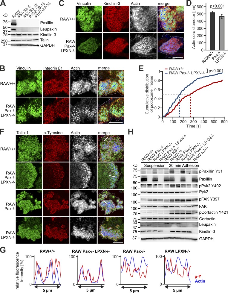
Figure 8.
Characterization of podosomes from paxillin/leupaxin double deficient RAW cells. (A) Western blot analyses of +/+ RAW cells and four different clones of paxillin/leupaxin dKO RAW cells for their expression of kindlin-3 and talin. (B and C) IF stainings of +/+ and paxillin/leupaxin dKO RAW cells for vinculin (green), integrin β1 (red, B), kindin-3 (red, C) and actin (white/blue in merge). (D) Diameter of the podosome actin cores in +/+ and paxillin/leupaxin dKO RAW cells. 10 actin cores in two regions of five to eight cells were measured in each experiment. n = 9/7. (E) Control RAW cells and different clones of paxillin/leupaxin dKO RAW cells were transfected with LifeAct-GFP and imaged at a spinning disc microscope for 10 min with a 15-s time interval. The cumulative distribution of these measurements is shown. 20 podosomes were measured per cell. At least four cells were analyzed in each of six dishes. (F) IF stainings for talin (green), tyrosine-phosphorylated proteins (red) and actin (white/blue in merge) on +/+, paxillin/leupaxin dKO, paxillin−/− and leupaxin−/− RAW cells. (G) Fluorescence intensity profiles of actin (blue) and phospho-tyrosine (p-Y; red) through three actin cores (indicated by the white lines in E) of +/+, paxillin/leupaxin dKO, paxillin−/−, and leupaxin−/− RAW cells. (H) Western blot analyses of the phosphorylation status of paxillin, Pyk2, FAK and cortactin in +/+, paxillin−/−, leupaxin−/−, K3−/− and paxillin/leupaxin dKO RAW cells kept in suspension or adherent to fibronectin. Scale bars, 10 µm. Dotted white lines mark cell borders.

