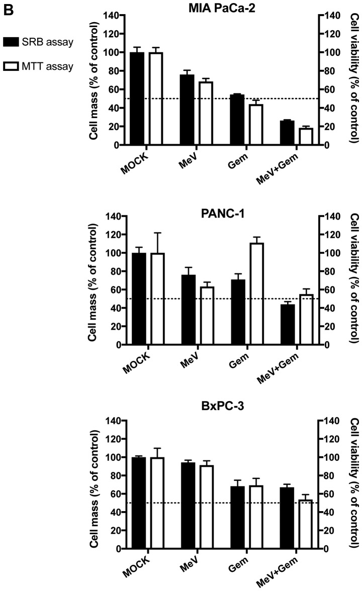
Figure 2.
Chemovirotherapy employing Gem together with the oncolytic measles vaccine virus in three pancreatic cancer cell lines. (A) Setting: Tumor cells were infected with MeV-SCD (here denoted as MeV) at 24 h after plating. Add-on of Gem was performed at 3 hpi when medium was changed. The total incubation time of virus was 72 h. (B) Cell viability was measured using two different assays [SRB (black bars) and MTT (white bars) assays, respectively] and normalized to an uninfected (MOCK-treated) control (set to 100% cell viability). MOIs of MeV and Gem concentrations were chosen at low enough levels to reduce tumor cell masses <50% when used as a single compound. When used in combination, the remaining tumor cell mass was found to be <5% in all three tumor cell lines. For MeV, MOIs of 0.4 (MIA PaCa-2), 0.075 (PANC-1) and 0.125 (BxPC-3) were chosen, respectively. For Gem, concentrations of 0.03 µM (MIA PaCa-2), 0.075 µM (PANC-1) and 0.02 µM (BxPC-3) were used, respectively. Data are presented as the mean ± standard deviation of three independent experiments. GEM, gemcitabine; hpi, hours post infection; MOIs, multiplicities of infection; MeV, measles vaccine virus; SRB, Sulforhodamin B.


