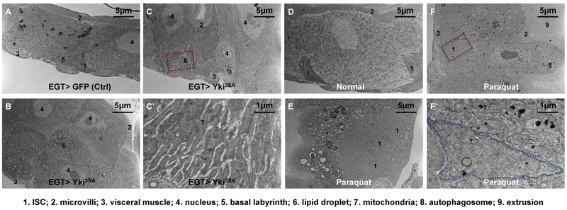
Figure 2.
Electron micrographs of midguts under pathological conditions. (A) The pseudostratified epithelium of midguts expressing GFP in ISCs for 4d with the EGT (esgGal4, UAS-GFP, tubGal80ts) expression driver. (B) The multi-layered epithelium of midguts expressing Yki3SA in ISCs for 4d. (C) A normal EC stretching from the basement membrane to the lumen found in a section of midgut Yki tumor. The magnified view of its extended basal labyrinth is shown in (C′). (D) Midgut epithelium of wild type (genotype: w1118) flies fed on normal food. (E) EC vacuolation and ISC expansion in the midgut epithelium of wild type flies fed on food containing 2 mM paraquat for 2d. Note that under tissue damage conditions the ISCs might no longer be enriched with free ribosomes, but they could be recognized by their cell boundary, small nuclei, and basal localization. (F) Deposition of electron-dense materials in the mitochondria and autophagosomes of ECs, but not in ISCs. Magnified view of an ISC and its neighboring EC is shown in (F′).

