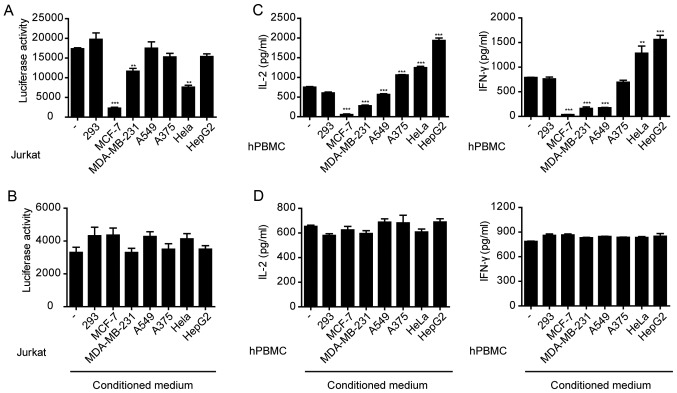
Figure 1.
Immunosuppression by cancer cells. (A) Jurkat cells transfected with PGL3-NFAT-TA-Luciferase plasmid were co-cultured with various cancer cell lines and stimulated with anti-CD3 (1 µg/ml) and anti-CD28 (1 µg/ml). Luciferase activity was measured 24 h after stimulation. **P<0.01, ***P<0.001 vs. control (−). (B) Jurkat cells transfected with PGL3-NFAT-TA-Luciferase plasmid were cultured in various cancer cell-conditioned media and stimulated with anti-CD3 (1 µg/ml) and anti-CD28 (1 µg/ml). Luciferase activity was measured 24 h after stimulation. (C) Human PBMCs were co-cultured with various cancer cell lines and then stimulated with anti-CD3 (1 µg/ml) and anti-CD28 (1 µg/ml). **P<0.01, ***P<0.001 vs. control (−). (D) Human PBMCs cultured in various cancer cell-conditioned media were stimulated with anti-CD3 (1 µg/ml) and anti-CD28 (1 µg/ml). hPBMC, human peripheral blood mononuclear cells; IL-2, interleukin-2; IFN-γ, interferon-γ.

