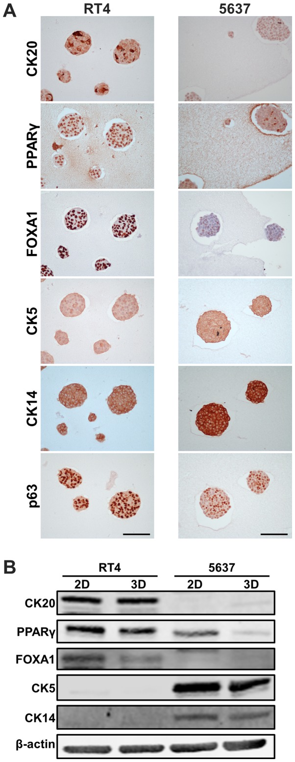
Figure 2.
Luminal/basal markers in RT4- and 5637-derived MCSs. (A) Immunohistochemical staining of RT4- and 5637-deried MCSs using antibodies against luminal markers (CK20, PPARγ and FOXA1) and basal markers (CK5, CK14 and p63). Scale bar, 100 µm. (B) Western blotting shows protein levels of luminal and basal markers in RT4 and 5637 cells cultured in 2D adherent culture and as MCSs in 3D suspension culture. β-actin was used as a loading control. MCSs, multicellular spheroids; MCSs, multicellular spheroids; CK, cytokeratin; PPARγ, peroxisome proliferator-activated receptor γ; FOXA1, forkhead box A1.

