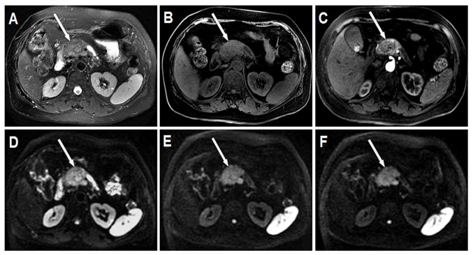
Figure 3.
Grade 2 pNEN located in the head of the pancreas (white arrow). The pancreatic body and tail exhibit atrophy and the pancreatic duct exhibits dilation. (A) Axial T2W MRI with fat suppression demonstrates pNEN with heterogeneous moderate hyperintensity and a relatively clear boundary. (B) Axial LAVA Flex water phase image displays pNEN with heterogeneous moderate hypointensity and an obscure boundary. (C) Axial arterial phase image demonstrates pNEN with mild heterogeneous enhancement. (D) Axial DWI with a b-value of 200s/mm2 demonstrates pNEN with heterogeneous hyperintensity and an obscure boundary. (E) Axial DWI with a b-value of 1,500s/mm2 demonstrates pNEN with heterogeneous hyperintensity and a clear boundary. (F) Axial DWI with a b-value of 2,000 s/mm2 demonstrates pNEN with heterogeneous hyperintensity and a clear boundary. pNEN, pancreatic neuroendocrine neoplasm; LAVA, liver acquisition with volume acceleration; DWI, diffusion weighted imaging; T2W, fast spin echo transverse relaxation time-weighted.

