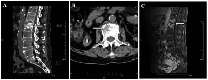
Figure 2.
Computed tomography and magnetic resonance imaging images of the lumbar vertebral body in a 54-year-old female patient on December 30, 2016. The patient did not receive any treatment after the first examination. (A) On computed tomography, a small-spot lamellar low-density shadow and a hardening edge around the L2 vertebral body in radiographs in the sagittal plane were visible, with no significant compression in vertebral height. (B) In the transverse plane of the vertebral body, the destruction of the vertebrae did not encroach on the spinal canal, and there was no compression of the dura or spinal cord. (C) On magnetic resonance imaging (visual field, 40×40×40 cm; layer thickness, 5 mm; layer spacing, 3 mm; b, 800 sec/mm2), patchy long T1 and T2 signals, and a high short T inversion recovery signal of the L1 and L2 vertebral body in the sagittal plane were observed, without invasion of the disk. Small grid, 1 cm. The lesion is indicated by white arrows.

