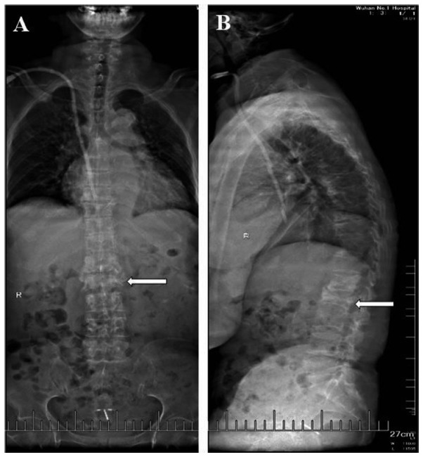
Figure 3.
X-ray of the lumbar vertebral body in a 54-year-old female patient on June 26, 2017. The patient did not receive any treatment. (A) Anteroposterior X-ray in the coronal plane and (B) lateral X-ray in the sagittal plane. Destruction of the vertebral bone, narrowing of the intervertebral space and partial necrosis was observed. Small grid, 1 cm. The lesion is indicated by white arrows.

