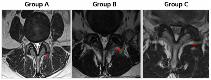
Figure 1.
Grading of facet joint osteoarthritis in representative T2-weighted axial MRI images of the lumbar spine. Group A, narrowing of the lumbar facet joint space and presence of a small osteophyte; group B, narrowing of the joint space, moderate osteophytes and/or subchondral erosions; and group C, narrowing of the joint space, large osteophytes and subchondral erosion/cysts are apparent. Red arrows indicate the facet joints.

