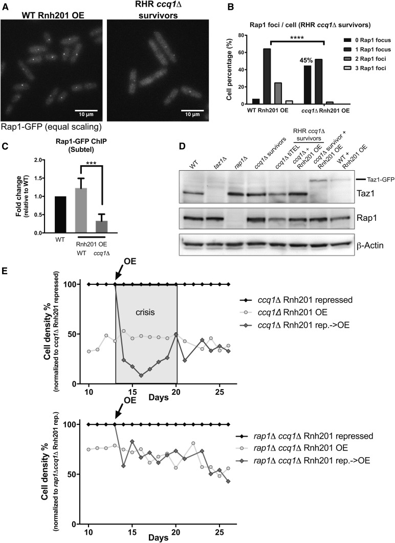
Figure 5.
Loss of Rap1 promotes formation of RHR ccq1Δ survivors in the context of constitutive Rnh201 overexpression. (A) Fluorescence imaging of Rap1-GFP reveals decreased recruitment to the telomere upon long-term induction of Rnh201 in ccq1Δ survivor cells. Fluorescence micrographs of WT or ccq1Δ Rap1-GFP survivor cells after long-term Rnh201 overexpression (RHR ccq1Δ survivors). Maximum intensity projections obtained as described in the Materials and Methods. Bar, 10 μm (B) Quantification of the number of Rap1-GFP foci after long-term induction of Rnh201 in the WT and ccq1Δ backgrounds. At least 200 cells were analyzed. P < 0.0001 by K-S test. (C) The loss of Rap1 association with telomeres is also observed upon Rnh201 overexpression by chromatin immunoprecipitation-qPCR. Normalized to WT cells and plotted as the mean with SD from three biological replicates. *** P = 0.0002 by Student’s t-test. (D) Taz1 levels increase in the absence of Rap1 and in all varieties of ccq1Δ survivors while Rap1 levels are unaffected. Whole cell extracts were separated on SDS-PAGE, blotted, and probed with antibodies to Taz1, Rap1, or β-actin (as a loading control). (E) Deletion of Rap1 prevents the growth crisis upon Rnh201 overexpression in ccq1Δ survivors. Serial culturing of either ccq1Δ (top) or rap1Δccq1Δ (bottom) Pnmt41-rnh201+ cells under constitutive repression (black), overexpression (light gray circles), or induced induction of overexpression (OE at day 13, black arrow; dark gray diamonds). Cell density is normalized to the Rnh201-repressed condition. The gray box indicates the period of telomere crisis upon overexpression of Rnh201 in ccq1Δ cells that is absent in rap1Δccq1Δ cells.

