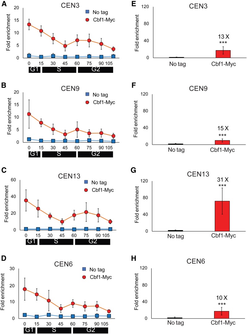
Figure 2.
Cbf1 binding to centromeres is cell cycle-regulated. (A–D) WT cells expressing Cbf1-MYC and growing at 24° were synchronized as described in Figure 1. Samples were removed at the indicated times for ChIP-qPCR analysis of Cbf1-MYC binding (red circles) to (A) CEN3, (B) CEN9, (C) CEN13, and (D) CEN6. Scales are not the same on all panels. ChIP-qPCR was carried out in parallel on a no-tag strain (blue squares). (E–H) show levels of Cbf1-MYC binding in 30°-grown asynchronous cells (red bars) and in a no-tag strain (blue bars). Cbf1-Myc binding was determined at (E) CEN3, (F) CEN9, (G) CEN13, and (H) CEN6. Values are the average of Cbf1-MYC binding in three independent colonies. The fold changes in the levels of binding on a given CEN of Cbf1-MYC relative to WT are shown above the red bars. Error bars and statistical analyses are as in Figure 1. ChIP, chromatin immunoprecipitation; qPCR, quantitative PCR; WT, wild-type.

