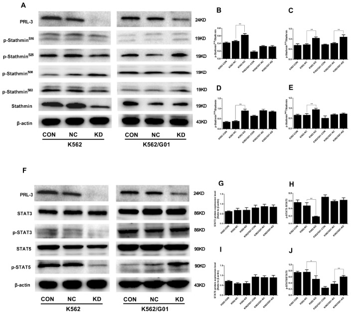
Figure 5.

The protein phosphorylation of stathmin and STATs signaling expressed in K562 and K562/G01 cells after PRL-3-silencing. (A) Stathmin, the four stathmin-serine sites, and β-actin expression were detected by western blot. (B–E) Quantification of stathmin-phospho (Ser16, Ser25, Ser38, and Ser63) were normalized to stathmin. (F) STAT3, p-STAT3, STAT5, p-STAT5 and β-actin expression were detected by western blot. (G–J) Quantification of p-STAT3 and p-STAT5 were normalized to STAT3 and STAT5, respectively. (*P<0.05, **P<0.01, vs. NC group).
