Figure 6.
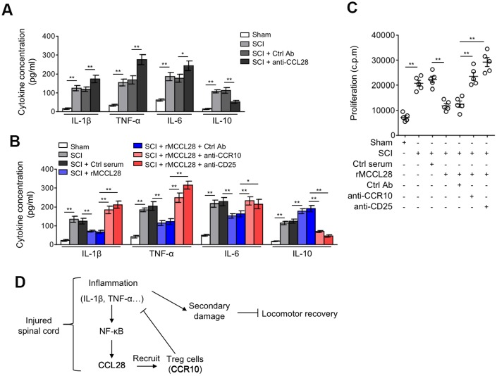
Treg cells mediate immune suppression in the spinal cord after SCI. (A) Mice were treated as in Figure 5A. ELISA analysis of cytokine concentration in the spinal cord at 7 days after sham or SCI surgery (n=5). (B, C) Mice were treated as in Figure 5B. (B) The cytokine concentration in the spinal cord at 7 days after sham or SCI surgery were determined as in (A) (n=5). (C) The proliferation rate of effector T cells was determined by [3H]-thymidine incorporation analysis (n=5). c.p.m., counts per minute of incorporated [3H]-thymidine. Data are mean ± SD. The statistical analysis was performed using Student’s t-test. **, P<0.01; *, P<0.05. (D) A brief schematic model of this study. After SCI, inflammatory cells infiltrate into the spinal cord and secrete cytokines, including IL-1β and TNF-α, which promptly induces the production of CCL28 via NF-κB activation. Responding to increased CCL28 in the focal sites, CCR10-expressing Treg cells are recruited and then exert their immune suppressive activities, restricting the inflammation to a controllable extent along with the time consumed. Owing to the activity of Treg cells recruited by CCL28, the local levels of IL-1β and TNF-α are decreased, thereby in turn relieving the stimulative effect on CCL28 upregulation, through this negative feed-back loop, CCL28 functions to suppress inflammation, reduce secondary damage and promotes locomotor recovery after SCI.

