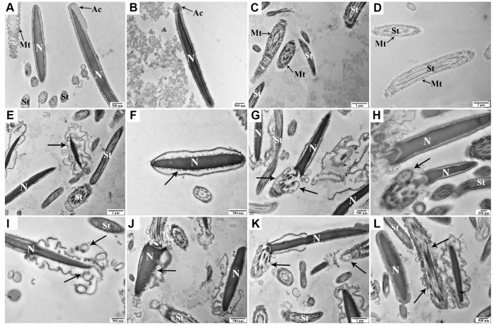
Figure 2.
Transmission electron microscopy images of morphologic changes in goat spermatozoa at 0 h (A–D), 48 h (E–H), and 96 h (I-L) of liquid storage. (A, B) Spermatozoa exhibited the normal ultrastructure. (C, D) Morphologically intact mitochondria. (E) Membrane blebbing. (F) Nuclear envelope defects. (G, H) Mitochondrial swelling, vacuolation, and deformity. (I) Membrane blebbing and apoptotic body formation. (J) Nuclear fragmentation. (K, L) Mitochondrial swelling, vacuolation, deletion, and disordered arrangement. Nuclear (N), acrosome (Ac), spermatozoa tail (St), and mitochondria (Mt). Scale bar = 1 μm (C, D, E, K), 500 nm (A, B, F, G, I, J, L), and 200 nm (H).

