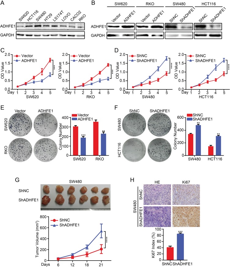
Figure 2.
Exogenous ADHFE1 knockdown promotes the proliferation of CRC cells, and the upregulation of ADHFE1 inhibits the proliferation of CRC cells. (A) Expression analyses of ADHFE1 protein in different CRC cells using western. GAPDH was used as a loading control. (B) Western blot analysis of the overexpression and knockdown of ADHFE1 in CRC cell lines. GAPDH was used as a loading control. (C and D) CCK8 analyses of the CRC cell proliferation with ADHFE1 overexpression or knockdown. (E and F) Colony formation analyses of the CRC cell proliferation with ADHFE1 overexpression or knockdown. (G) The xenograft models were generated after injecting SW480/ShNC and SW480/ShADHFE1 cells in nude mice (n = 6/group). The tumor volumes were measured on the indicated days. The data points represent the mean tumor volumes ± SD. (H) The sections of tumor were subjected to H&E staining or IHC staining using an antibody against Ki-67. Error bars represent the means ± SD from three independent experiments. **p<0.01, ***p<0.001, ****p<0.0001.

