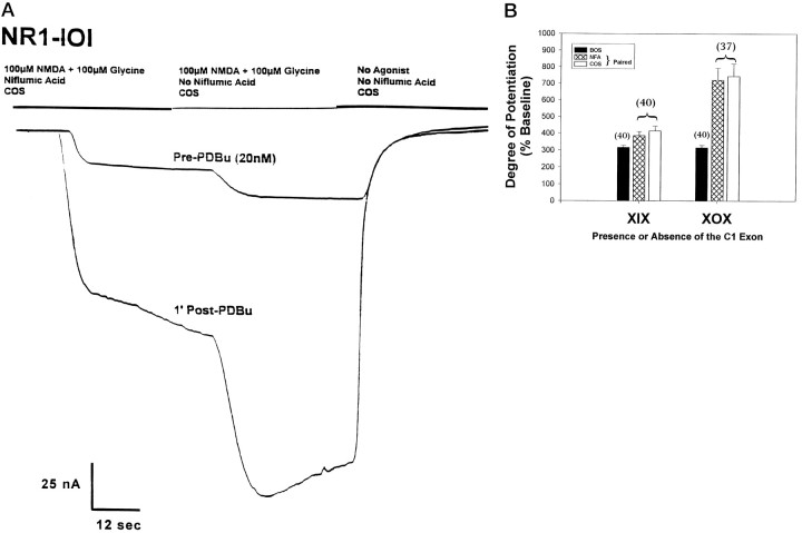
Fig. 4.
Niflumic acid (NFA), a blocker ofICl(Ca), has no effect on the high level of current potentiation by PKC observed in NR1-X0X variants.A, The traces represent whole-cell currents recorded at −80 mV from oocytes injected with NR1-101. The experimental design is represented by the bars at the top of the current traces. Before a trial was run, the test cell was incubated in COS containing 200 μm NFA for 1 min. Agonist (100 μm NMDA, 100 μm glycine) containing 200 μm NFA was applied for 40 sec followed by an application of agonist alone (i.e., no NFA present) for 40 sec followed by a COS wash (no agonist, no NFA). Once a stable baseline was reached (Pre-PDBu), the cell was incubated in 20 nmPDBu for 8 min. The cell was then washed for 1 min in COS containing NFA, and currents were evoked in the manner described. Thereafter, currents were elicited every 2 min to generate a time course from which the peak level of current potentiation was determined. In the example shown here, the peak potentiation was reached after the first COS/NFA wash (1′ Post-PDBu). The current shown potentiated from 19.8 to 100.1 nA (506%) in the presence of NFA and from 33.2 to 172.1 nA (518%) after NFA washout. B, Experiments were performed on all the splice variants (∼8 cells each) as described inA. The data were then grouped according to the presence or absence of the C1 exon, and the mean peak current potentiation was plotted. Numbers in parentheses indicate the number of cells used to generate the data. The open bracket is used to indicate that the NFA data and the COS data are collected from the same cell. The barium data (black bar) are included from the data set in Figure 2B for comparison.

