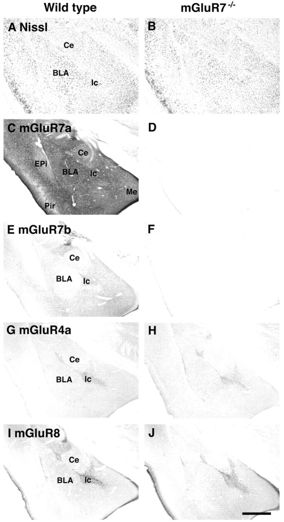
Fig. 2.

Histological and immunohistochemical analyses. Light microscope images of coronal sections are shown. No obvious change in the morphology of the amygdala was detected with Nissl staining of the mGluR7−/− knockout mouse as compared with the wild-type control (A,B). Moderate to intense immunostaining of mGluR7a and weak immunostaining of mGluR7b were observed in the amygdala of wild-type mice, but these immunostainings totally disappeared in sections of mGluR7−/− knockout mice (C–F). No obvious change in the patterns and extents of mGluR4a and mGluR8 immunostainings was detected between wild-type and mGluR7−/− knockout mice (G–J). BLA, Basolateral amygdaloid nucleus; Ce, central amygdaloid nucleus; EPi, endpiriform nucleus; Ic, intercalated nucleus; Me, medial amygdaloid nucleus;Pir, piriform cortex. Scale bar, 500 μm.
