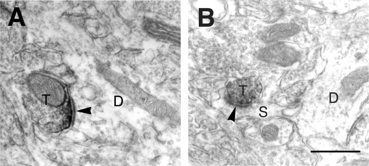
Fig. 3.
Immunoelectron-microscopic analysis. Electron micrographs showing immunoreactivity for mGluR7a in the BLA. Immunoreactive products for mGluR7a were accumulated along presynaptic membrane specialization in axon terminals (T), making asymmetrical synapses (arrowheads) on dendrites (D inA) and spines (S in B). Scale bar, 0.5 μm.

