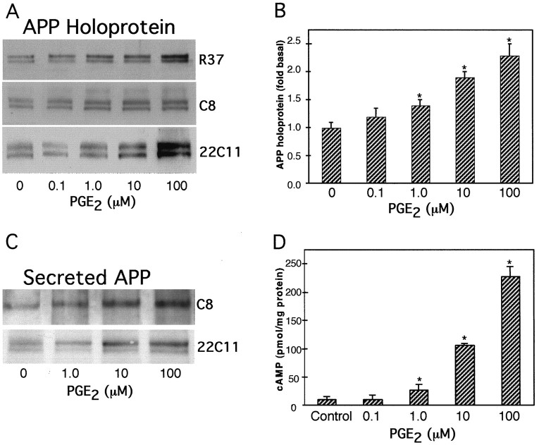
Fig. 1.
Effect of PGE2 on cellular and secreted APP and on cAMP production in cultured astrocytes.A, Representative immunoblots show that 0.1, 1, 10, and 100 μm PGE2 treatment for 24 hr stimulated dose-dependent increases in cellular APP holoprotein. The levels of APP holoprotein as measured by mAb 22C11, antisera R37, or antisera R98 did not differ significantly. B, The graph represents the means and SEM of APP holoprotein levels stimulated by different concentrations of PGE2 (*p < 0.05). Densitometric analysis of APP levels using mAb 22C11 (n = 2), antiserum R37 (n = 2), or antiserum R98 (n = 1) were expressed as arbitrary values and normalized to the levels obtained from untreated, control cells. C, Representative immunoblots showing the levels of APP released into the media, as detected by mAb 22C11 or antiserum C8. Both immunoblots revealed increased amounts of secreted APP after PGE2 treatment for 24 hr compared with control cells. Similar results were obtained from a subsequent experiment.D, Dose-dependent increases in cellular cAMP levels obtained by PGE2 treatment of astrocytes. Graph represents means and SEM from a representative experiment performed on duplicate dishes (*p < 0.05).

