
Fig. 1.
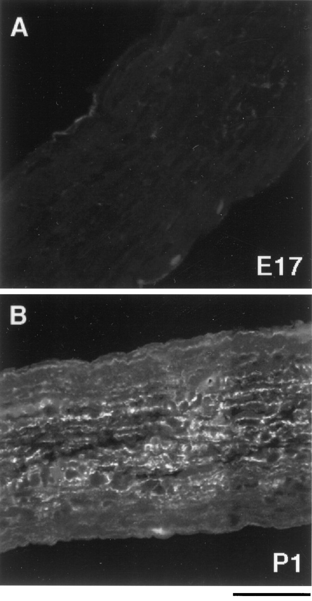
The appearance of GFAP immunoreactivity in the developing rat optic nerve. Optic nerves from embryonic day 17 (A) and postnatal day 1 (B) rats were fixed, sectioned longitudinally, and stained with an anti-GFAP antibody. GFAP expression was scarcely detected in E17 optic nerve (A) but was found throughout theP1 optic nerve (B). Scale bar, 50 μm.