
Fig. 6.
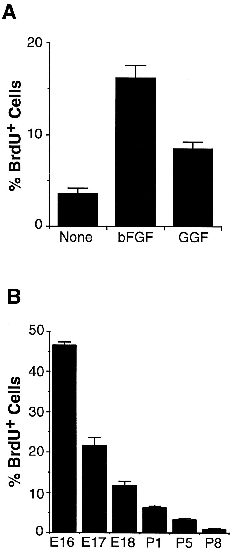
Proliferation of astrocyte precursor cells.A, Purified APCs were cultured in bFGFfor 1 hr, followed by washing and culture in serum-free B-S medium containing CPT-cAMP and the indicated peptide factors. After 4 d, the cells were incubated with BrdU for 2 hr and stained by an anti-BrdU antibody. The percentage ofBrdU+ APCs over total APCs was counted. The results represent means ± SD of three wells of a single experiment and were confirmed by at least three separate experiments. B, Proliferation of astrocyte lineage cellsin vivo is shown. Two hours after an intraperitoneal injection of BrdU, optic nerves of various ages were sectioned and stained by a monoclonal anti-BrdU antibody and a polyclonal anti-Pax2 antiserum. At each age, the percentage of Pax2+ cells that was alsoBrdU+ was determined. The results are means ± SEM of six sections from three different animals in each age group.