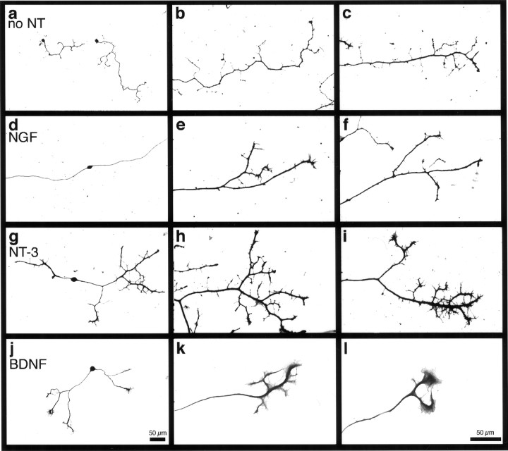
Fig. 5.
NGF, NT-3, andBDNF mediate different morphological responses.a, d, g, j, Low-power photomicrographs (scale bar in j).b, c, e, f,h, i, k, l, High-power photomicrographs (scale bar in l). For visualizing morphologies of individual cells, neurons fromBax−/− mice were plated at low density (500 neurons per well) and grown for 72 hr in vitro without neurotrophins or in the presence of 50 ng/ml NGF,NT-3, or BDNF. a–c, Representative Bax−/− neurons cultured in the absence of neurotrophins (no NT). The neurons were unipolar and extended numerous short branches from the primary axon along its entire length. d–f, TypicalNGF-responsive Bax−/− neurons cultured in the presence of NGF. These neurons were bipolar and extended long, relatively unbranched axons. NGFtreatment suppressed the extension of short branches close to the cell soma. Typically, a few thick caliber branches were present along the distal portion of the axon. g–i, Typical NT-3-responsive Bax−/− neurons in the presence ofNT-3. NT-3 also appeared to suppress branching near the soma, although the primary axons of neurons treated with NT-3 were shorter than were those ofNGF-treated neurons. NT-3–responsive neurons almost invariably exhibited elaborate branching at the distal ends of their axons. j–l, RepresentativeBax−/− neurons depicting the characteristic morphological appearance of a subset of neurons that respond toBDNF. Note the prominent lathellipodia.

