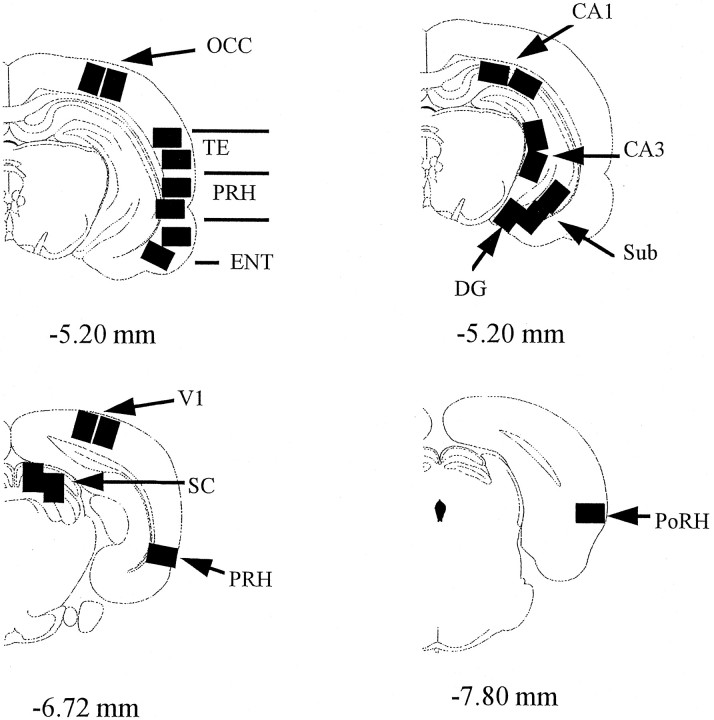
Fig. 2.
Diagrams of coronal sections indicating areas sampled. The distance (in millimeters) of the sections from bregma is indicated (Paxinos and Watson, 1986). So that processing of the different areas could be done in parallel, only the illustrated brain levels were sampled. One area was sampled for the dentate gyrus and postrhinal cortex, three areas for perirhinal cortex, and two for all other regions. The significance or nonsignificance of any of the reported effects was not dependent on these differences in the number of sampling points. CA1, CA3, Hippocampal subfields; DG, dentate gyrus; ENT, entorhinal cortex; OCC, occipital cortex;PoRH, postrhinal cortex; PRH, perirhinal cortex; SC, superior colliculus; TE, area TE of the inferotemporal cortex; V1, primary visual cortex; VLG, ventral lateral geniculate nucleus of the thalamus. The locations of PRH and PoRH are modeled after Burwell et al. (1995).

