Fig. 2.
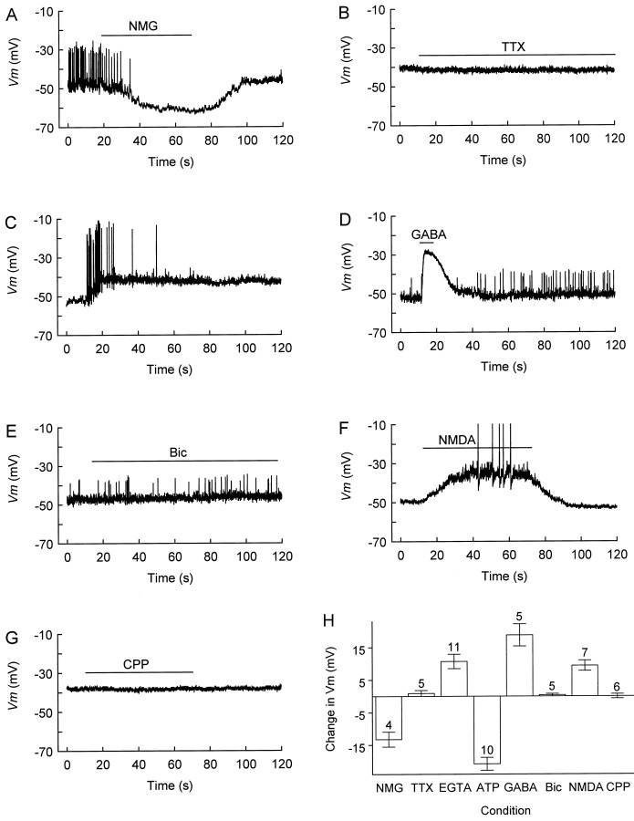
Modulation of CR cell resting potential by various agents. A, Local substitution of extracellular Na+ ions byN-methyl-d-glucamine (NMG) hyperpolarizes membrane potential (Vm) and blocks spontaneous firing. For unknown reasons, firing was not restored immediately on return to control (Na+-rich) medium but reappeared 3.8 min later. B, Tetrodotoxin (TTX; 1 μm) fails to change Vm.C, After 7 min of whole-cell recording with 10 mm EGTA in the pipette, a burst of action potentials steadily depolarizes Vm. D,GABA (100 μm) transiently depolarizesVm, producing a shunting inhibition of firing. E, Bicuculline fails to affectVm or firing. F, NMDA (100 μm) depolarizes Vm to spike threshold (the spikes are truncated). G, CPP does not affect Vm. H, Summary of the effects of the drugs that were tested. Zero is taken as the initial resting potential; a negative value indicates hyperpolarization, and a positive value corresponds to depolarization. The numbers above the error bars indicate the number of cells tested. A, D, E, Perforated-patch recording.B, C, F, G, Whole-cell recording. All tests were performed in regular Ringer’s (plus 2 mmMg2+; no glycine was added). Drugs were applied for the duration indicated by bars above the traces.

