
Fig. 8.
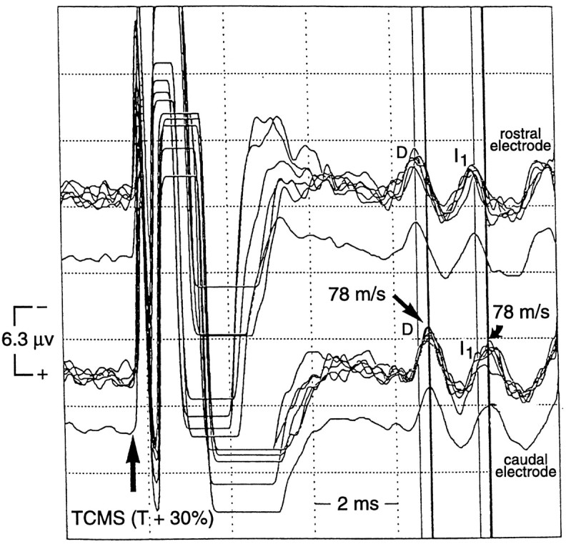
Descending SCEPs recorded from epidural electrodes at two different spinal cord levels (near Th8) afterTCMS at SCEP T + 30% in one alert subject. The epidural recording electrodes were rostrocaudally separated by 2.0 cm, and each was referenced to a surface electrode (G2) over the Th8 vertebrae. The high bandpass filter was 500 Hz (bandpass, 500 Hz to 5 kHz) on both channels to reduce muscle artifacts contributed by the surface electrode. The SCEP waves recorded from the rostral epidural electrode had shorter latencies than did those recorded from the caudal electrode. The spinal cord conduction velocity of the D and I1 wave was 78 m/sec.