Fig. 2.

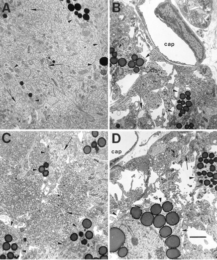
The typical ultrastructure of the NL is shown in electron micrographs of coronal sections from 1 week postsurgical intact control (A), 1 week postlesion (B), 4 week postlesion (C), and 4 week chronic hyponatremic postlesion (D) animals. A, The intact NL is characterized by densely packed neurosecretory axons, frequently filled with neurosecretory vesicles, interspersed with glial (pituicyte) processes (arrowheads) that often contain osmiophilic lipid inclusions. Smaller axonal profiles lacking vesicles are identifiable by the presence of neurofilaments (arrows).B, By 1 week after the hypothalamic lesion, the number of axons is dramatically reduced, and the amount of extracellular space is substantially increased. C, At 4 weeks after the lesion, these degenerative changes have been largely reversed as collateral sprouting has returned the number of axon profiles to near normal levels. D, In contrast, the axon population does not recover in rats maintained under CH during the 4 week postlesion interval, and a substantial amount of extracellular space remains.cap, Capillary lumen. Scale bar, 2 μm.
