Fig. 4.
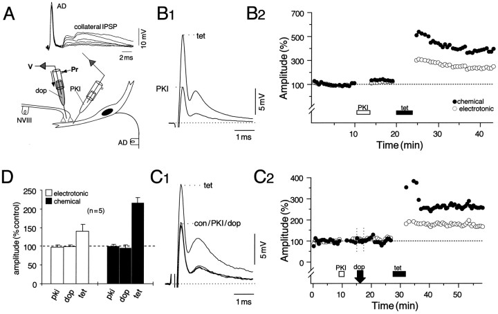
LTP does not require the activation of cAMP-dependent protein kinase (PKA). A, Schematic of the experimental arrangement for intradendritic pressure injections (below) with superimposed consecutive traces of the passively conducted antidromic action potentials (AD) and the chloride-dependent collateral IPSP recorded during the injections (above). The transient increase in the IPSP indicates successful pressure injection of a solution containing 900 μm of the cAMP-dependent protein kinase inhibitor (PKI). B1, B2, NVIII tetanization in the presence of PKI still produces a robust and persistent potentiation. B1, Superimposed averaged (n = 15) intradendritic recordings of the M-cell response to NVIII stimulation after injection of the inhibitor (PKI) and ∼10 min after tetanization (tet). B2, Plot of normalized amplitudes (ordinate) of the electrotonic coupling potential (open circles) and the chemical EPSP (filled circles) versus time (abscissa), with PKI and tetanus applied during the indicated periods. In this and subsequent figures, 100% equals the mean of all of the corresponding control (con) values, and each point is an average of at least 15 response amplitudes evoked at a stimulus rate of 0.5 Hz. C1,C2, To ensure that the injected peptide was active, we applied dopamine (dop) to the dendrite through an extracellular microelectrode before tetanization. Shown are averages of the response to NVIII stimulation (C1) and the time course of changes in the electrotonic coupling and chemical PSP (C2) after the indicated sequence of manipulations. Note that, as in B2, the chemical EPSP is potentiated more than the coupling potential. D, Pooled data (n = 5) demonstrating that the inhibitor blocks dopamine-evoked potentiation yet spares tetanus-induced LTP. The bar plots represent changes in the mean amplitude (% control) and SEM (error bars) of the coupling potential (open bars) and the chemical EPSP (filled bars) after PKI, dopamine, and tetanization. Measurement samples in this and subsequent bar plots were taken from the steady-state regions of the time courses, typically 10–15 min after a perturbation. After tetanization, the mean (μ) potentiations (±SEM) of the chemical EPSP [μ(c) = 116 ± 14%] are consistently larger than the coupling potential [μ(e) = 40 ± 18%] in this series, but not statistically different from controls (p > 0.05; n = 7).

