Fig. 5.
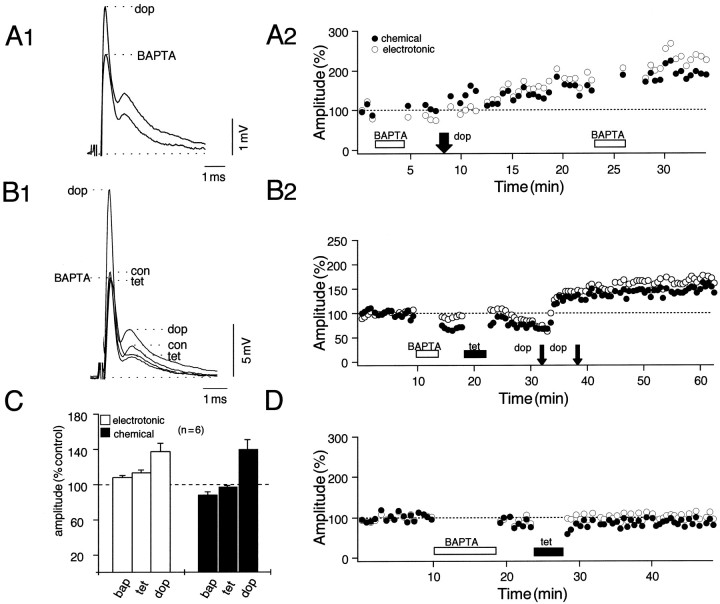
Postsynaptic BAPTA injections reveal the differential role of calcium in the induction of tetanus and dopamine-evoked potentiations. The experimental arrangement is as in Figure 1A. A1, A2, Data from an experiment in which dopamine (dop) was applied locally after intracellular injection of BAPTA (20 mm).A1, Superimposed averaged (n = 15) intradendritic recordings of the NVIII response in the presence of BAPTA before and after the application of dopamine. A2, Plot of normalized amplitudes of the electrotonic coupling potential (open circles) and the chemical EPSP (filled circles) as a function of time, with BAPTA and dopamine applied during the periods indicated. A second BAPTA injection ∼15 min after dopamine confirmed the calcium-independent effects of the amine. B1, B2, Further evidence for the separation of intracellular pathways. Ineffectiveness of the tetanus in the presence of the chelator confirmed the successful injection of the latter, whereas dopamine still produced a potentiation. C, Bar plots from six experiments indicating changes in the mean (±SEM) amplitude (% control) of the coupling potential (e;open bars) and the chemical EPSP (c;filled bars) that follow, in order, BAPTA, tetanus, and dopamine. The mean (μ) potentiations (±SEM) evoked by dopamine after tetanization in this series [μ(e) = 24 ± 15%;μ(c) = 47 ± 14%] did not differ statistically from control experiments (p > 0.05; n = 6). Note that there is a small reduction in the chemical EPSP (μ = 12 ± 6%) immediately after an injection of BAPTA. D, Plot from a control experiment in which a single tetanus was applied to NVIII shortly after BAPTA injection.

