
Fig. 6.
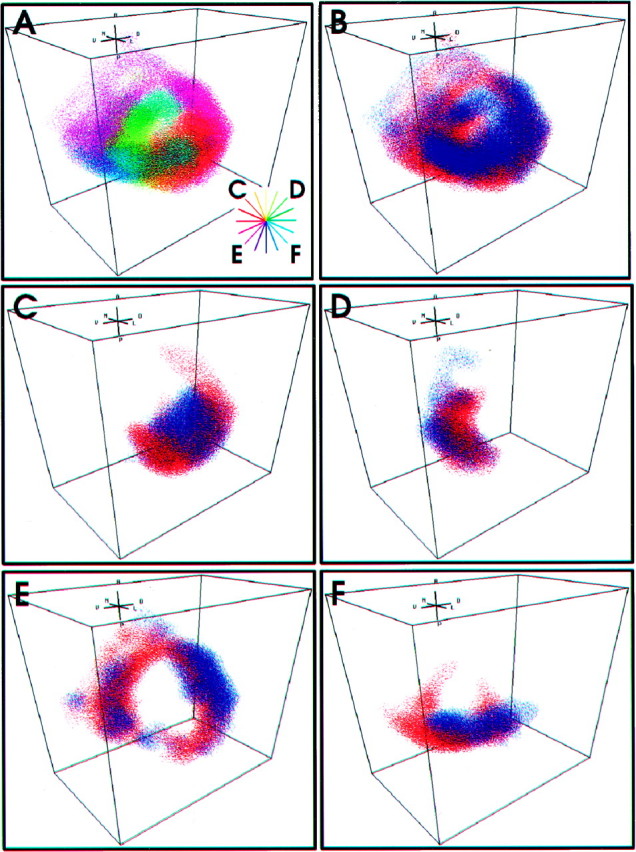
Anatomical segregation between two functional representations. A, Functional representation of stimulus direction by L and M afferents, color coded according to peak directional tuning with respect to body coordinates.Inset, The color wheel corresponds to the peak directional tuning in body coordinates of the subsets of afferents shown in C–F. B, Functional representation of stimulus frequency, represented by the L (red) and M (blue) afferent arborizations. C–F, Arborization patterns of subsets of L and M afferents tuned to different air current directions. C, Three L afferents and three M afferents with peak directional sensitivities near −45°. D, Three L afferents and three M afferents with peak directional sensitivities near 45°. E, Three L afferents and three M afferents with peak directional sensitivities near −135°.F, Three L afferents and three M afferents with peak directional sensitivities near 135°.