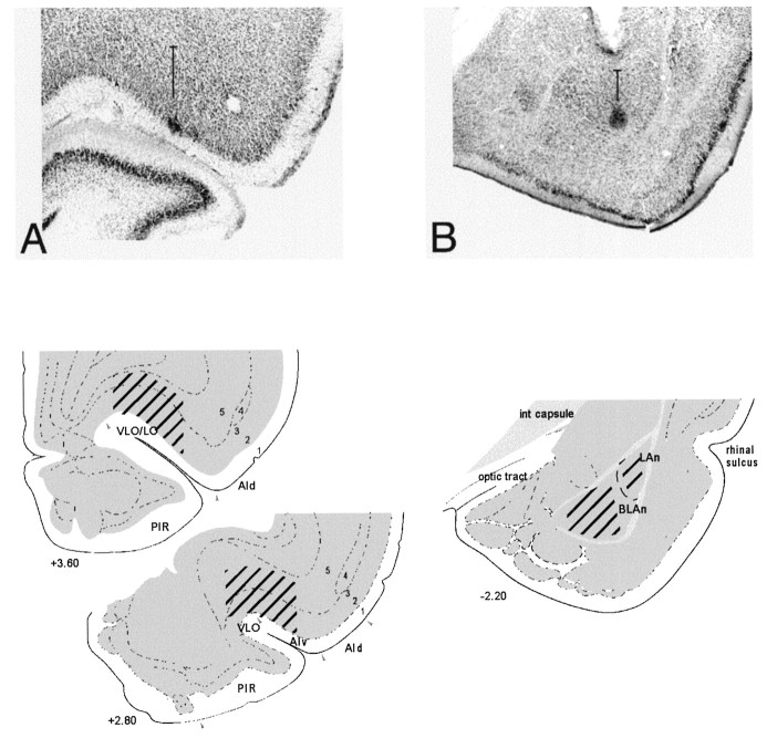
Fig. 2.
Electrode recording sites. Photomicrographs of histological sections showing the reconstruction of recording sites in representative subjects in OFC (A) and ABL (B). In each photomicrograph, a vertical line represents the dorsoventral range along the electrode track from which neurons were recorded in the case shown. Below each photomicrograph is a drawing that shows the approximate area in which recordings were obtained in each group. The OFC encompasses the orbital regions and agranular insular cortex. Recordings were localized to ventrolateral and lateral orbital regions (VLO/LO) and ventral agranular insular cortex (AIv) in the four rats in the OFC group. Recordings were localized to the basolateral nucleus in three of the rats in the ABL group (pictured in photomicrograph and as BLAn in drawing) and lateral nucleus in the fourth rat (LAn). (Drawings adapted from Swanson, 1992; photomicrographs adapted from Schoenbaum et al., 1998.)

