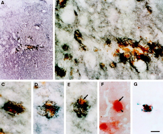
Fig. 9.

Photomicrographs showing DAT-positive sprouting dopaminergic fibers growing toward and around hemosiderin-filled wound macrophages. A, Low-magnification image showing bundles of dark DAT-positive fibers sprouting in the periwound margin. B, High-magnification image showing sprouting fibers approaching and growing around hemosiderin-containing macrophages. C–E, Higher magnification images showing the intimate association of sprouting dopaminergic fibers to a number of individual hemosiderin-filled (arrow) wound macrophages. F, Photomicrograph demonstrating that hemosiderin (arrow) is located within macrophage cytoplasm (arrowhead) (NSE stain). G, NSE-positive macrophage containing prussian blue deposits of iron-containing hemosiderin. Scale bar:A, 375 μm; B, 25 μm;C–G, 10 μm.
