
Fig. 3.
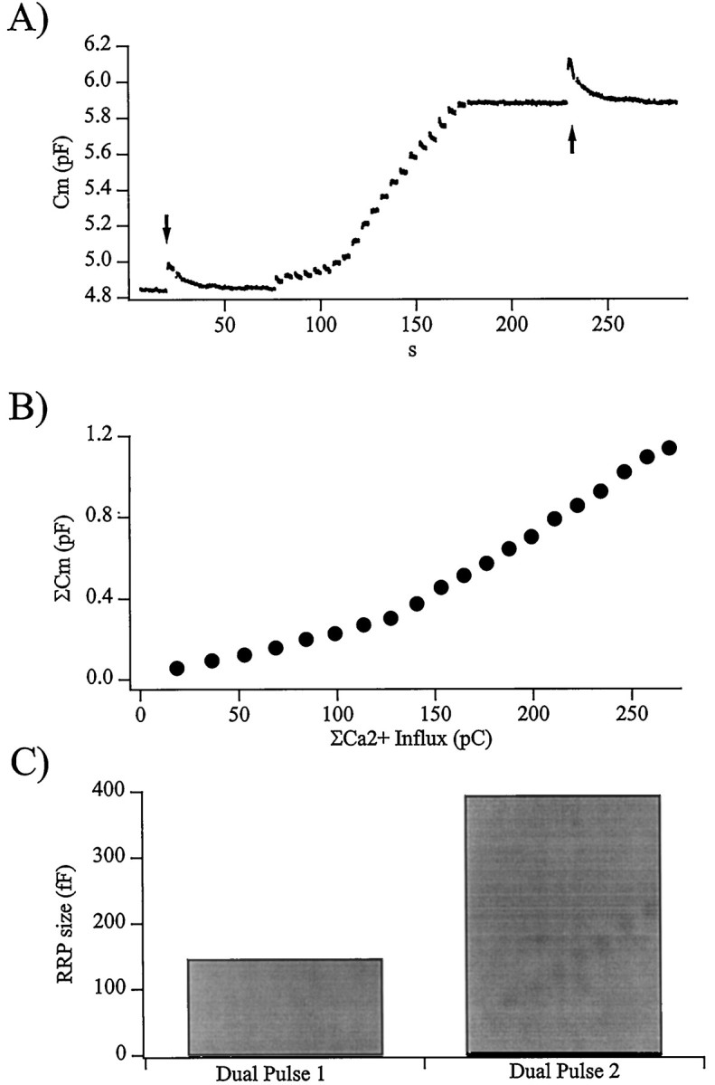
The activity-dependent facilitation is caused by a larger RRP of vesicles. A, Plotted is the membrane capacitance recorded from a cell stimulated by a pulse protocol designed to measure the activity-dependent changes in the number of vesicles in the readily releasable pool. The initial size of the RRP was determined through the use of a dual-pulse protocol (arrow). The RRP was then allowed to completely recover before the cell was stimulated by a single train of depolarizations as described in the legend to Figure 1. Again, after complete recovery to its steady state, the size of the RRP was measured by a dual-pulse stimulation protocol (arrow). The values R for the first and second dual-pulse stimuli in this cell were 0.28 and 0.57.B, As in Figures 1 and 2, the integrated evoked cell capacitance increase is plotted against total evoked Ca2+ influx, showing a shift in secretory efficiency during the train. The total evoked increase in Cm was 1136 fF in response to 269 pC total Ca2+ influx, an exceptionally high increase of exocytotic efficiency during the train. This cell was chosen for presentation because both dual-pulse stimuli resulted in an adequate amount of exocytotic depression for an accurate estimate of the RRP. C, In response to the train of depolarizations, the size of the RRP grew from a pretrain value of 146 fF to the increased value of 393 fF.