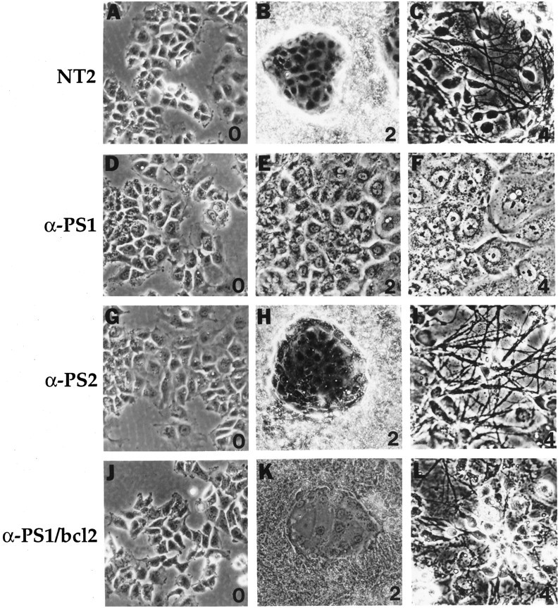
Fig. 1.

Morphological changes of NT2 cells and transfected NT2 cells during neuronal differentiation. A–C, NT2 cells in basal conditions (0) and after2 and 4 weeks of RA treatment. Note that the 2 week RA culture shows a characteristic bimodal pattern with large groups of clumped multilayered cells surrounding small islands of epitheloid cells. The latter cells are flat and apparently nondividing and contrast sharply to the rapidly dividing neuronal progenitor cells. The 4 week RA culture shows a dense neuritic network extending from neurons that overlay the epitheloid cell layer. D–F, Representative anti-PS1 cell line in basal culture conditions (0) and after 2 and4 weeks of RA induction. In comparison to normal NT2 cells, the anti-PS1 cells consist exclusively of flat epitheloid cells, even after 4 weeks of RA treatment, without any evidence of neuronal differentiation. The neuritic processes are conspicuously absent. Note that these cells are strikingly similar to the epitheloid cells in normal NT2 cells. G–I, Representative anti-PS2 cell line in basal culture conditions (0) and after2 and 4 weeks of RA induction. The anti-PS2 cells resemble control NT2 cells at basal and RA-treated conditions. These cultures differentiate normally into neurons after 4 weeks. J–L, Representative anti-PS1 cell line transfected with bcl-2 cDNA (anti-PS1/bcl-2 cells) at basal culture conditions (0) and after 2 and4 weeks of RA treatment. In comparison with anti-PS1 cells, the anti-PS1/bcl-2 cells resemble control NT2 cells showing characteristic morphology and differentiate into neurons. Magnification, 240×.
