
Fig. 4.
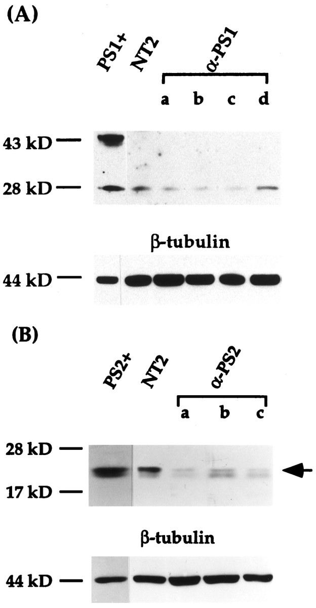
Immunoblot analysis of PS1 and PS2 proteins in transfected NT2 cell lines. NT2 cells were transfected with either antisense-oriented PS1 or PS2 cDNA. Equal amounts of total proteins (30 μg) were separated by SDS-PAGE, transferred to nitrocellulose membranes, and incubated with either J27 polyclonal antibody to PS1 or PS2 monoclonal antibody. A, The amount of N-terminal PS1 proteolytic fragment is reduced in the differentiation-defective anti-PS1 cell lines (a–c) compared with that of control NT2 cell line but not from a PS1 antisense transfected cell line with normal differentiation (d). Note that in addition to the ∼28 kDa N-terminal proteolytic fragment, full-length PS1 protein (∼43 kDa) is present in a Chinese hamster ovary (CHO) cell line overexpressing PS1 (PS1+) but not in any of the NT2 cell lines. Bottom panel, The filter was stripped and incubated with β-tubulin antibody for normalization of the amount of protein loaded in each lane. B, PS2 expression of anti-PS2 cell lines. The ∼20 kDa PS2 C-terminal fragment (arrow) was detected in a CHO cell line stably transfected with PS2 (PS2+), control NT2 cells, and representative anti-PS2 cell lines. PS2 is reduced to different levels in the anti-PS2 cells (a–c), but all the cell lines differentiated into neurons by RA. In this autoradiogram, there is a faint band just below the PS2 C-terminal fragment that is present throughout the samples to varying levels (see Fig.2B). Because this band is neither increased in overexpressing cells nor decreased in antisense transfected cells, it appears to be a cross-reactive species. Bottom panel, The filter was incubated with β-tubulin antibody after antibody stripping.