
Fig. 3.
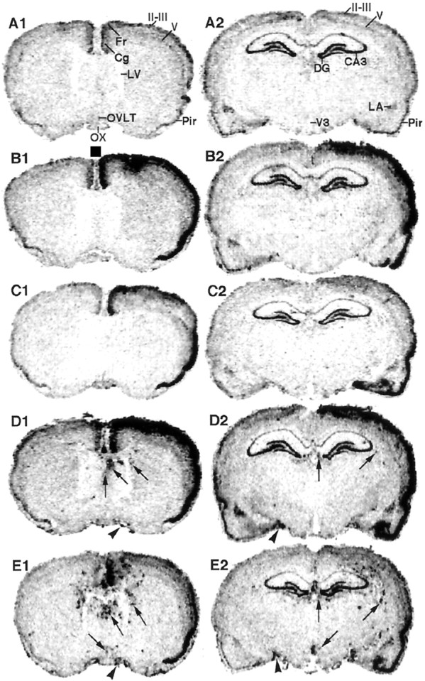
Macroautoradiographic images of COX-2 mRNA signals in coronal sections of the rat brain. InB1–E2, the sections were arranged so that the side in which intracerebroventricular injection was made was shown on the right. In the brain sections of untreated rats (A1, A2), the COX-2 mRNA signal was observed in the cingulate/frontal cortices, layer II-III and V of the neocortex, piriform cortex, lateral amygdala, and hippocampus. The mRNA signals were markedly enhanced in one side of the cerebral cortex where the injection of either saline (B1, C2) or LPS (D1, E2) had been made. Until 1.5 hr after LPS injection (D1, D2), but not saline injection, the spot-like COX-2 mRNA signals appeared in the brain parenchyma near the cerebral ventricles (arrows) and in the subarachnoidal space (arrowheads). Until 3.5 hr after LPS injection (E1, E2), the spot-like COX-2 mRNA signals markedly increased in the number and intensity. OX, Optic chiasma; Cg, cingulate cortex; Fr, frontal cortex;Pir, piriform cortex;II-III, the second and third layers of the neocortex; V, the fifth layer of the neocortex;LV, lateral ventricle; V3, third ventricle; DG, dentate gyrus; CA3, field CA3-in Ammon’s horn; LA, lateral amygdala.