
Fig. 9.
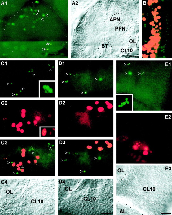
Coincidence of apoptotic cell death and cell proliferation in the lobster olfactory brain as demonstrated by double labeling with the TUNEL assay (green, to label apoptotic cells) and phos H3 (red ororange, to label mitotic cells). A, Whole mount of a brain at E25%, photomontage. A1, TUNEL assay, numerous nuclei are labeled in the protocerebrum (single arrowheads) but only a few in the deutocerebrum (double arrowheads). A2, same specimen under bright-field illumination. B, Whole mount of an eyestalk anlage at E25%; the proliferation zone of the medulla externa (orange) is flanked by two bands of apoptotic cells (green). C, Whole mount of the olfactory lobe (OL) and cell cluster 10 (CL10) of a stage 7 juvenile; see C4 for labels. C1, TUNEL assay, arrowheads point to labeled nuclei;inset, higher magnification of a TUNEL-labeled fragmented nucleus that displays a typical apoptotic morphology.C2, phos H3, mitotic cells in PD10;inset, higher magnification of nuclei in prophase or metaphase (left) and in anaphase (right).C3, double exposure of C1 andC2. C4, Bright-field image of the same specimen. D, Vibratome section, brain of a 1.5-year-old juvenile; see D4 for labels. D1, TUNEL assay, arrowheads point to labeled nuclei.D2, Phos H3, mitotic cells in PD10. D3, Double exposure of D1 and D2.D4, Bright-field image of the same specimen.E, Vibratome section, brain of a 7-year-old adult; seeE3 for labels. E1, TUNEL assay,arrowheads point to labeled nuclei;inset, higher magnification of a TUNEL-labeled fragmented nucleus that displays a typical apoptotic morphology.E2, Phos H3, mitotic cells in PD10. E3, Bright-field image of the same specimen. AL, Accessory lobe; APN, anterior protocerebral neuropil;CL10, cell cluster 10; OL, olfactory lobe; PPN, posterior protocerebral neuropil. Scale bars:A–E, 50 μm.