Fig. 4.
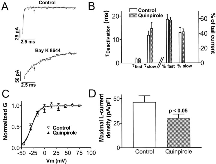
Chronic quinpirole treatment in vitro induces a long-lasting suppression of L-type Ca2+ channel current density without changing its functional properties. A, Illustration of the method of isolating L-channel tail current from total Ca2+channel current in melanotropes (see Results for details). Noisy traces are 10 msec portions of tail currents recorded at −50 mV after a step depolarization to +75 mV. The smooth traces are exponential curves fit to the currents. The time constants are 0.16 msec (monoexponential curve in top) and 2.5 and 21.9 msec (biexponential curve in bottom). Thearrows are placed at 2.4 msec after repolarization to −50 mV. B, Chronic quinpirole treatment does not alter L-channel deactivation properties. Data were obtained with biexponential curve-fitting analysis of Bay K 8644-slowed tail currents in control and quinpirole-treated cells. Bars on theleft and right halves of the graph correspond to the left and right y-axes, respectively.C, Chronic quinpirole treatment does not alter the voltage-dependence of activation of L-channels. Normalized conductance (G) versus step depolarization potential (Vm) data for control and quinpirole-treated cells are fitted with Boltzmann equations (smooth curves). D, Maximal L-current density in melanotropes cultured for 6–10 d in control or quinpirole-containing media. Maximal L-current density values were 46.3 ± 6.6 pA/pF in control cells and 30.1 ± 4.1 pA/pF in quinpirole-treated cells. Data in B–D come from 9 control cells and 13 quinpirole-treated cells.

