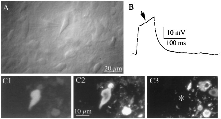
Fig. 1.
Identification of OT and VP neurons recorded from hypothalamic slices. A, Photomicrograph of a SON neuron visualized with infrared–differential interference contrast videomicroscopy. Note the recording pipette attached to the neuron.B, The presence of a strong transient outward rectification (arrow) to depolarizing current injection (50 pA) is characteristic of magnocellular neurons. C, Example of an immunochemically identified VP neuron. The neuron was filled with biocytin and immunochemically labeled for VP– and OT–neurophysin by double immunofluorescence. In C1, The recorded neuron is visualized with amino-methylcoumarin-conjugated avidin.C2, The recorded neuron is positively labeled with VP–neurophysin immunoreactivity visualized by fluorescein-conjugated secondary antibody. C3, OT–neurophysin immunoreactivity visualized by tetramethylrhodamine-conjugated secondary antibody. The recorded neuron (*) was negative.

