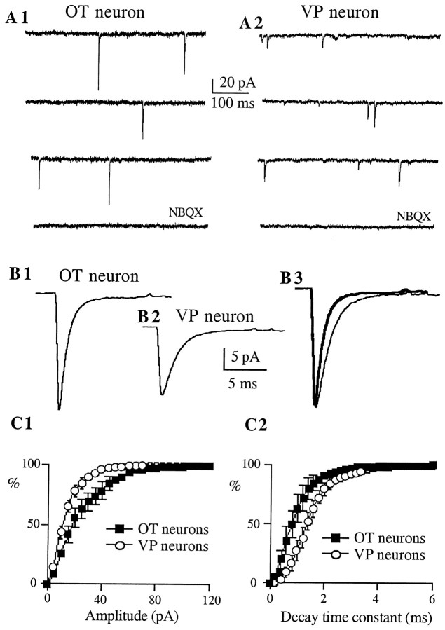
Fig. 3.
AMPA mEPSCs of OT neurons show larger amplitude and faster decay kinetics as compared with VP neurons.A, Representative AMPA mEPSCs obtained from immunoidentified OT (A1) and VP (A2) neurons. Synaptic activity was recorded in the presence of bicuculline (20 μm) and ±APV (100 μm), at a holding potential of −70 mV. These synaptic events were blocked by NBQX (10 μm). B, Average of 317 mEPSCs from the same OT neuron (B1) and average of 486 mEPSCs from the same VP neuron (B2). Note the larger amplitude of the averaged mEPSC obtained in the OT neuron. In B3, both responses were scaled to the same (larger) peak amplitude to facilitate the comparison of their decay times. Note the faster decay kinetics of the averaged mEPSC of the OT neuron (thick line trace).C, Averaged cumulative distribution histograms of the amplitude (C1) and decay time constant (C2) of AMPA mEPSCs obtained from eight OT (squares) and VP (circles) neurons. The amplitude and the decay time constant distributions were significantly different between cell types (p < 0.0001 in both cases; Kolmogorov–Smirnov Test).

